

Dermoscopy Mole Evaluation
Expert mole assessment with high-resolution dermoscopy
Dermoscopy allows our dermatologists to visualise subsurface skin structures using polarised or contact light, dramatically improving the accuracy of mole and lesion assessment compared to the naked eye — giving patients clarity about which lesions are safe and which require removal.
Regular dermoscopic mole mapping is recommended for patients with multiple moles, a family history of melanoma or a history of sunburn — enabling early detection when skin cancers are most treatable.
Key Benefits
- High-accuracy evaluation of pigmented skin lesions
- Early detection of suspicious lesions
- Avoids unnecessary removal of benign moles
- Baseline digital documentation for future comparison
Is This Treatment Right For You?
- Patients with many moles or a family history of melanoma
- Those who notice a changing or new pigmented lesion
- Anyone wanting a baseline skin cancer screening assessment
What to Expect
Patient History
Personal and family skin cancer history and sun exposure are discussed.
Full Skin Examination
A systematic head-to-toe skin examination is conducted.
Dermoscopy Assessment
Suspicious lesions are examined in detail under dermoscopy.
Management Plan
Benign lesions are documented; suspicious lesions are biopsied or excised.
Recovery & Aftercare
No recovery for examination only; biopsy or excision sites heal within 5–14 days.
Frequently Asked Questions
How often should I have moles checked?
Annual mole checks are recommended for most patients; higher-risk patients may need 6-monthly reviews.
What happens if a suspicious mole is found?
The dermatologist will recommend a biopsy or excision; the tissue is sent for histopathological analysis to confirm the diagnosis.
Patients Also Explore
Our specialists offer a full range of complementary treatments across surgery, skin, hair and wellness.
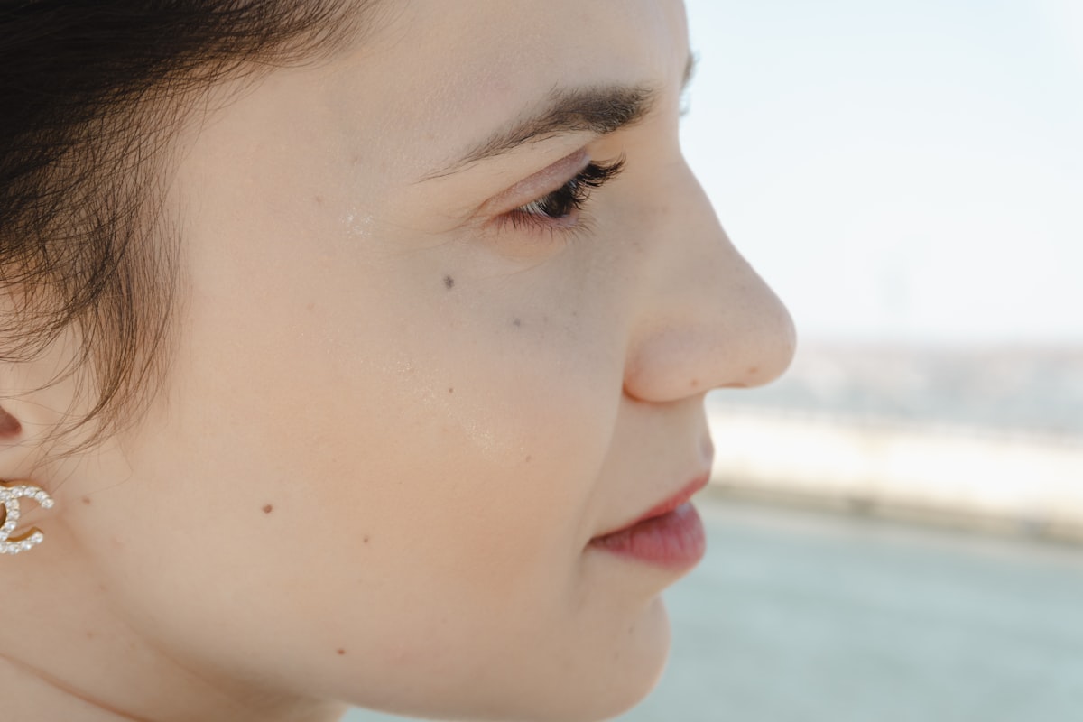
Rhinoplasty
Refine the shape, size or profile of your nose while preserving natural breathing function.
Learn More
Breast Augmentation
Enhance breast volume and shape using high-quality implants or fat transfer.
Learn More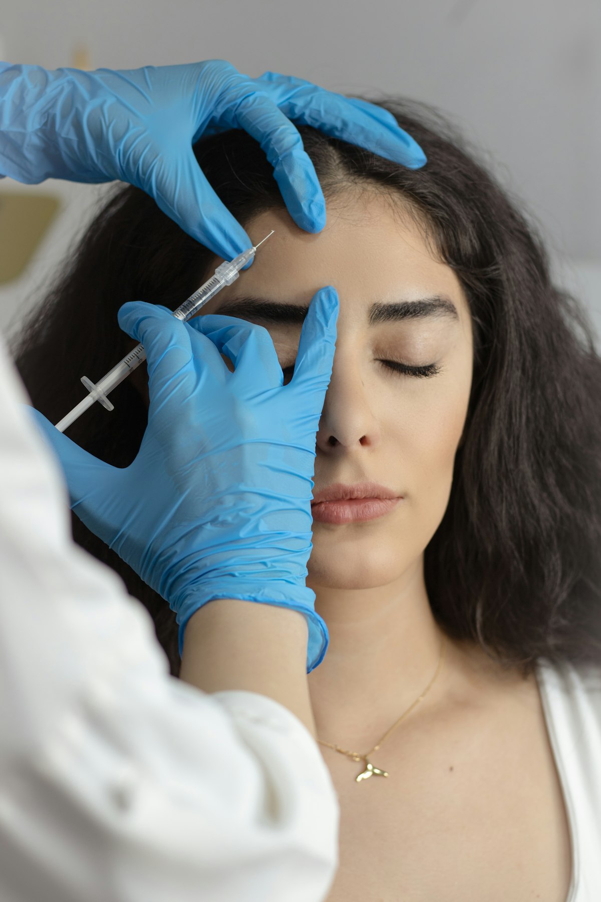
Botox
Relax the muscles that cause frown lines, crow's feet and forehead creases.
Learn MoreReady to Book Your Dermoscopy Mole Evaluation Consultation?
Speak to a specialist and get an honest, personalised recommendation.